Ocular Surface Squamous Neoplasia (OSSN) / Pterygium
Ocular Surface Squamous Neoplasia (OSSN) / Pterygium
The term ocular surface squamous neoplasia represents a spectrum of lesions ranging from simple dysplasia of the cornea and conjunctival to invasive squamous cell carcinoma. It can be subtle clinically and diagnosis difficult on clinical appearance alone, thus histological confirmation is often required which means obtaining a tissue sample, either via impression cytology or surgical excision.
When surgery is required, the pathology is excised and the area treated with cryotherapy (freezing). Sometimes, topical (drops) chemotherapy may be necessary.
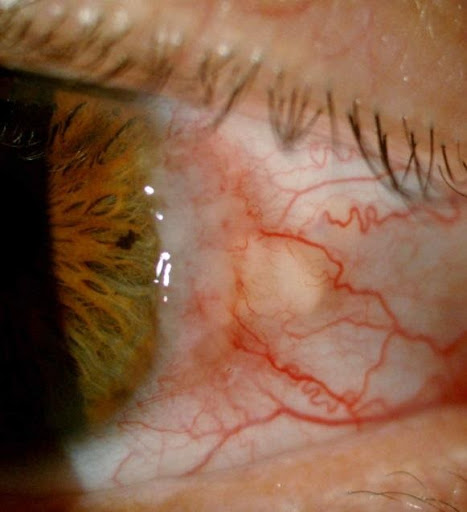
High grade conjunctival intra-epithelial neoplasia (CIN)

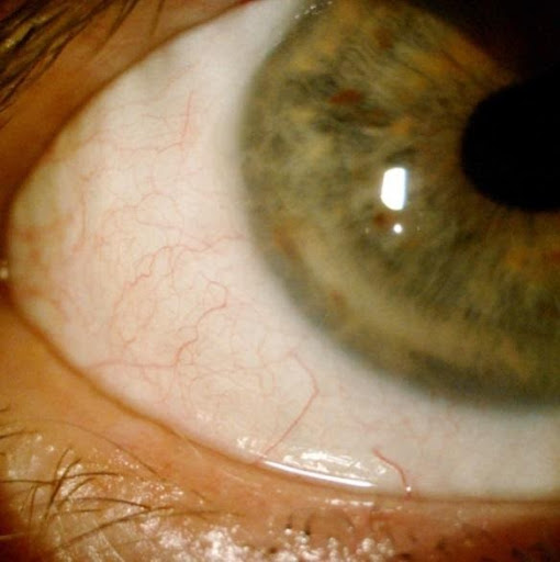
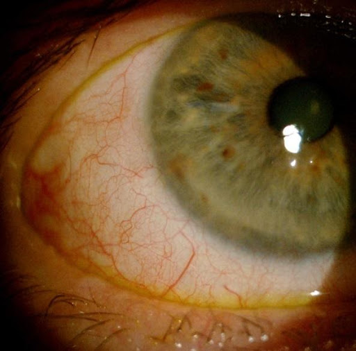
Mucoepidermoid carcinoma with overlying moderate to severe squamous dysplasia. Managed with wide margin excision with local cryotherapy and conjunctival graft.
Mucoepidermoid carcinoma post surgery, below left photo 2 years post-op, below right photo 4 years post-op
Pterygium is an abnormal overgrowth of conjunctival tissue that typically advances across corneal surface within the eyelid fissure, appearing as a triangular shaped lesion. It is a minor annoyance & cosmetic problem but may potentially cause visual impairment, scarring & restricted motility in recurrent lesions with multiple excisions.
It is often due to exposure to elements in the environment ie sun, wind, dust. For those who work outdoors, it is advisable to wear sunglasses to minimize the development of this problem.
Management is dependent on the size & site. Conservative approach includes reassurance, topical lubricants, vasoconstrictors, topical steroids with surgery reserved for advanced lesion, severe disfigurement or chronic irritation.
Pterygium surgery is performed under local anaesthetic as a day case procedure.
The pterygium is excised and a conjunctival graft is harvested from the same eye (often from the top of the eye) and either stitched or more commonly ‘glued’ onto the surgical site where the pterygium used to be.
The eye will be padded and be reviewed usually the next day. Recovery may take several days and the eye often may appear ‘red’ for some weeks.
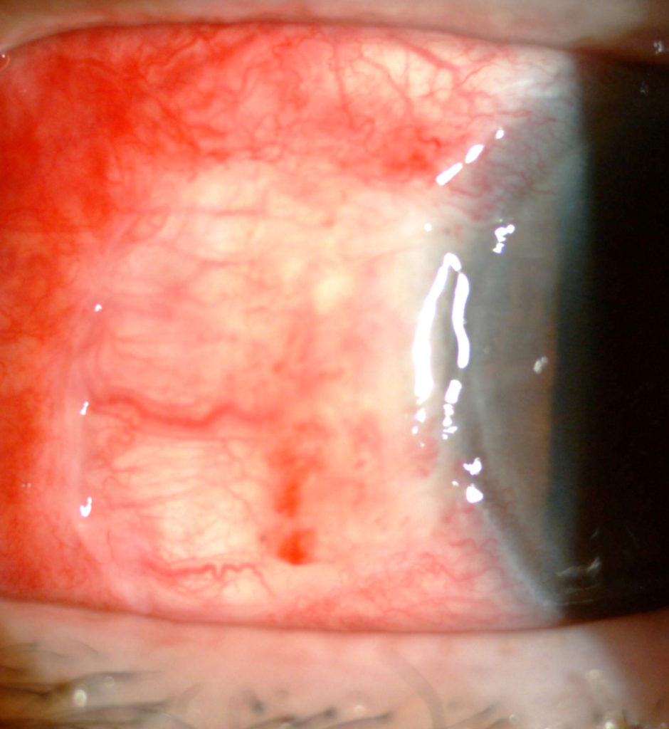
Day 1 post surgery
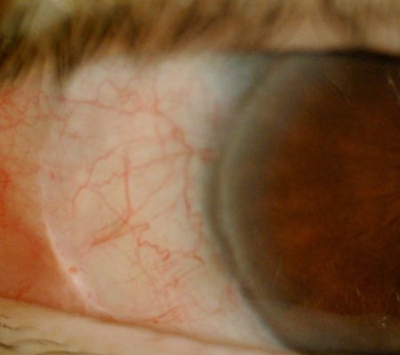
Week 6 post surgery
The link below provides general information from RANZCO
https://ranzco.edu/policies_and_guideli/pterygium/